February 2021
Should we really be irradiating so many platelets?
Dana V. Devine, PhD FCAHS
Chief Scientist, Canadian Blood Services and Director, University of British Columbia Centre for Blood Research
Irradiation of platelet concentrates is undertaken to prevent any residual white blood cells in the unit from being able to replicate. The process of irradiation causes lesions in DNA including single- and double-strand breaks, DNA-protein cross-links, and oxidization. Enough damage to DNA prevents cell replication. Most units of platelets are irradiated using a dose of 25 Gy (Gray) also expressed as 2500 rad or 25 Joule/kilogram. This is a measure of radiation and does not take into consideration the biological effect of the radiation dose.
Standards for red blood cells clearly recognize that irradiation causes damage to cell membranes of RBCs, shortening the allowable storage period post-irradiation. However, we have no specific storage limits on platelet concentrates that have been subjected to irradiation. With the advent of pathogen inactivation (PI) technologies, more attention has been paid to the exposure of platelets to irradiation as with PI, all products are exposed to ultraviolet radiation as part of the inactivation process. This has been determined to cause some damage to platelets and has led investigations of the impact of gamma irradiation on platelet concentrates. While the literature generally supports that gamma irradiation does not cause the same level of loss of platelet quality as the UV irradiation used in PI, there are reports of increased reactive oxygen species generation following gamma irradiation and associated acceleration of the development of the platelet storage lesion. Irradiated platelets have a tendency toward a lower pH and lower morphology score than non-irradiated platelets which reflects some damage caused by the radiation exposure.1,2 Our laboratory has observed similar changes to platelet quality parameters and platelet metabolism caused by irradiation of Canadian Blood Services platelets.
Others have reported some negative effects on transfusion efficacy caused by irradiation especially if performed early in the storage period.3,4,5
Thus, on balance, while it will be necessary to irradiate platelets for certain patients, in order to minimize damage to the platelets, this should be done close to the time of transfusion. Policies that have all platelet products irradiated upon arrival at the blood bank are likely to reduce the quality of those platelets and make the transfusion less efficacious.
- Marrocco C, D’Alessandro A, Girelli G, Zolla L. Proteomic analysis of platelets treated with gamma irradiation versus a commercial photochemical pathogen reduction technology. Transfusion 2013;53:1808–1820;
- Nodeh FM, Hosseini E, Ghasemzadeh M. The effect of gamma irradiation on platelet redox state during storage. Transfusion in press DOI: 10.1111/trf.16207
- Slichter SJ, Davis K, Enright H, et al. Factors affecting posttransfusion platelet increments, platelet refractoriness, and platelet transfusion intervals in thrombocytopenic patients. Blood 2005;105:4106–4114;
- Slichter SJ. Platelet transfusion therapy. Hematol Oncol Clin North Am. 2007;21:697–729;
- Julmy F, Ammann RA, Fontana S, et al. Transfusion efficacy of apheresis platelet concentrates irradiated at the day of transfusion is significantly superior compared to platelets irradiated in advance. Transfus Med Hemother. 2014;41:176–181
Registration now open:
April 14th: 16th Annual TM Education Web Conference

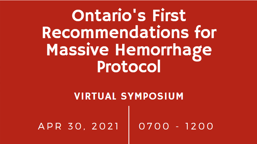
April 30th: Ontario’s First Recommendations for Massive Hemorrhage Protocol Virtual Symposium
Fetal – Neonatal Alloimmune Thrombocytopenia (FNAIT)
Gwen Clarke MD FRCPC
Associate Medical Director, Clinical Services
Canadian Blood Services
FNAIT is a rare and potentially serious cause of thrombocytopenia affecting a fetus or neonate in the first few days of life. This condition is an immune thrombocytopenia that occurs when maternal antiplatelet alloantibodies develop, pass through the placenta and bind to fetal platelets and megakaryocytes resulting in clearance of the platelets and decreased platelet production.
The pathophysiology of this disorder is like that of Hemolytic Disease of the Fetus and Newborn (HDFN). A mother is exposed to paternally inherited Human Platelet Antigens (HPA) expressed on fetal platelets or other tissues. In response she develops anti-HPA antibodies which then cross the placenta where they can bind to fetal platelets and megakaryocytes. Unlike HDFN, FNAIT may occur in a first pregnancy as alloimmunization may occur very early in gestation.
Following antibody binding, platelet clearance may lead to a range of clinical presentations from asymptomatic thrombocytopenia in the neonate, mild bleeding with petechiae or bruising, to severe bleeding and intracranial hemorrhage.
Human Platelet antigens are expressed on platelet glycoproteins. The clinically significant antigens are grouped into 11 groups of antithetical antigens called HPA1a/1b, HPA2a/2b, etc. The most common antibodies are those directed against HPA 1a, arising in pregnant women who have the HPA type 1b/1b while the fetal HPA type is 1a/1b. Other HPA antibodies may also be implicated, though less commonly. Ethnic background may also impact the HPA antigens most likely to cause alloimmunization.
While as many as 1 in 50 Caucasian women may have the HPA1b1b platelet type only 1 in 400 develop anti-HPA 1a antibodies. Even amongst those with antibodies, only 1/1500 develop severe thrombocytopenia with an even smaller number (~1/10 000) of pregnancies affected by intracranial hemorrhage. The development of antibodies and the severity of thrombocytopenia is linked to a particular HLA type (HLA-DRB3*01:01). Without this HLA type the formation of antibodies is uncommon. Testing for this HLA type amongst women known to have the HPA 1b1b type can help to determine the risk for severe thrombocytopenia.
Diagnosis of FNAIT is generally contemplated when neonatal thrombocytopenia or fetal or neonatal bleeding are identified in the absence of other common causes for thrombocytopenia. The diagnosis requires identification of anti-HPA antibodies in maternal plasma along with determining maternal and paternal HPA types through genotyping. The combination of maternal-paternal HPA incompatibility along with associated maternal anti-HPA antibodies and bleeding or thrombocytopenia in a fetus or neonate, is diagnostic.
For example, a maternal HPA 1b1b type together with a paternal HPA 1a1a type and maternal anti-HPA 1a antibodies would be a typical diagnostic pattern. The diagnosis is usually confirmed through use of a test that directly identifies reactivity between maternal antibodies in serum and paternal platelets such as a MAIPA (Monoclonal Antibody-specific Immobilization of Platelet Antigen).
Since there is no routine antenatal screening program for anti- platelet antibodies, usually antenatal treatment is offered to women with a previously affected fetus or neonate. Intravenous immune globulin is considered first line treatment for prevention of thrombocytopenia or bleeding due to FNAIT. There are recently published ICTMG guidelines outlining the selection of patients and the timing of treatment. (www.ictmg.org)
Following delivery platelet transfusions may be provided to thrombocytopenic neonates to maintain a platelet count greater than 30 X 109/L. The optimal platelet product for this indication is an HPA matched platelet (negative for the HPA antigen corresponding to the maternal antibody). If an HPA matched apheresis platelet is not immediately available random platelet pools will likely be effective in achieving hemostasis and should be provided until HPA matched platelets can be obtained.
Canadian Blood Services (CBS) routinely collects and maintains an inventory of HPA1b1b apheresis platelets to ensure availability for urgent use in neonates with FNAIT due to anti-HPA-1a. Donors with other HPA genotypes may be recruited to support platelet transfusions in neonates with FNAIT due to other antibodies.
In future, antenatal determination of the fetal HPA genotype may more routinely available through non-invasive perinatal testing from maternal plasma. Other future advances in this field may include immunoprophylaxis with anti-HPA1a antibodies (similar to RhIG therapy) for women known to have an HPA 1b1b type, in order to prevent alloimmunization.
Additional information related to the background, diagnosis and treatment of FNAIT along with a list of related references may be found at the CBS professional education website: https://profedu.blood.ca/en/transfusion/best-practices/testing-and-management-fetal-and-neonatal-alloimmune-thrombocytopenia
Upcoming Event:
February 25th: Fetal-Neonatal Alloimmune Thrombocytopenia (FNAIT) presented by Dr. Lani Lieberman and Ms. Lynette Beaudin.